
About syndrome
SUMMARY
THAT IS WHAT THE Gorlin syndrome? DEFINITION
DR Gorlin RJ, SURGEON-DENTIST AND geneticist, ITS COURSE
GENETIC CHARACTERISTICS OF Basal Cell Nevus Syndrome
A / GENERAL
B / GENE INVOLVED IN THE Basal Cell Nevus Syndrome
1. General
2. Theory of the double event
3. transduction Way Patched / sonic hedgehog
Actors transduction pathway patched / Sonic Hedgehog
The transduction pathway of patched operating / Sonic Hedgehog
Signaling pathway Patched / Hedgehog Sonic
Types of mutations and location
GENERAL EVENTS IN DETAIL, THE Basal Cell Nevus Syndrome
1 / EVENTS DERMATOLOGICALS
A / Naevi and basal cell carcinomas General
B / palmoplantar hyperkeratosis General
C / Dermatological events
2 / SKELETON EVENTS
A / brain calcifications envelopes
B / Abnormal skull shape
C / rib abnormalities
D / vertebral anomalies
E / Abnormal bone of the hand and foot
F / Other skeletal manifestations
3 / EYE EVENTS
A / Congenital malformations of the globe
B / Tumor globe process
4 / EVENTS NEUROLOGIC
A / Méduloblastomes Statistics
B / Other neurological manifestations
5 / OTHER GENERAL EVENTS
A / ovarian fibroids
B / heart Fibroids
C / hypogonadism in men:
6 / EVENTS ODONTOSTOMATOLOGIQUES
A / The odontogenic keratocysts
B / Other events odontostomatologiques
a / Inclusions or dental malposition
b / mandibular prognathism
c / lip and palate clefts
d / paresthesia
e / The améloblastome
THAT IS WHAT THE Gorlin syndrome? DEFINITION
Gorlin syndrome, also known as the basal cell nevus syndrome (NBC) is an inherited disease characterized by a set of developmental abnormalities and a predisposition to various cancers.
The prevalence is estimated at between 1/57 and 1/256 000 000, with a ratio male / female 1: 1.Clinical manifestations include the presence of numerous basal cell carcinomas (BCC), odontogenic keratocysts of the jaws, hyperkeratosis of palms and soles, skeletal abnormalities, intracranial ectopic calcifications and facial dysmorphism (macrocephaly, cleft lip-palato and severe eye anomalies).Intellectual deficit is observed in 5% of patients. Basal cell carcinomas (from papules having the color of the skin to ulcerative plates with a diameter ranging from 1 to 10 mm) are usually located on the face, back and chest. The number of basal cell carcinomas ranged from a few to several thousand.Skeletal abnormalities (affecting the shape of the ribs, vertebrae and skull) are frequent. May also occur eye, genito-urinary and cardiovascular disorders. Among patients with Gorlin syndrome, 5-10% develop medulloblastoma, which is a probable cause of early death.
The syndrome is caused by mutations in the gene PTCH1 and is transmitted as an autosomal dominant with complete penetrance and variable expressivity. The clinical diagnosis is based on specific criteria.The diagnosis is confirmed by research mutations. Genetic counseling is mandatory. Antenatal diagnosis is possible by ultrasonography and DNA analysis of fetal cells obtained by amniocentesis or chorionic villus sampling. The main differential diagnosis includes Bazex syndrome, multiple trichoépithélium and Muir-Torre syndrome syndrome (see these terms). The support requires a multidisciplinary approach.
Keratocysts are removed by surgical resection. In the case of basal cell carcinomas, surgery is indicated when the number of lesions is limited. Other possible treatments are laser ablation, photodynamic therapy, and local chemotherapy.
Radiation therapy should be avoided. The analogues of vitamin A may play a preventive role against development of new CBC. Life expectancy is not significantly altered but complications can result in significant morbidity. Regular monitoring by a multidisciplinary team (dermatologists, neurologists and odontologists) is essential. Patients should avoid excessive exposure to UV rays.
Publisher (s) expert (s)
DR Gorlin RJ, CHRIRURGIEN- DENTIST AND geneticist, ITS COURSE

Robert James Gorlin was born January 11, 1923 in Hudson, New York and died of lymphoma August 29, 2006 in Minneapolis, Minnesota at the age of 83. Joined the army during World War II, he then studied dentistry at the University of Washington where he graduated in 1947. He continued his education by obtaining a Master of Chemistry at the University of State of Iowa. After numerous academic positions, in 1956 he obtained a position as Professor and Director of the Oral Pathology Division at the University of Minnesota, where he remained until his death.
His career took a turn in 1940 when Gorlin discovers a book of Helen Curth dermatologist dealing with achantosis nigricans. This experience will help transform Gorlin in "syndromologiste". The achantosis nigricans is a skin disease characterized by vegetating papillary hypertrophy and pigmentation localized mainly in the armpits, neck and genital-femoral regions where the skin appears rough, thickened and squared. This dermatological condition has malignant forms characterized by an association with gastric adenocarcinoma. It is a dermatological condition that affects other body systems. This discovery has captivated Gorlin which then asked if certain systemic diseases could present oral manifestations. Gorlin became internationally known for his work on oral manifestations of many syndromes. Throughout his career, Robert J. Gorlin and his colleagues studied and named about 100 syndromes and participated in the isolation and characterization of genes responsible for about half of them.
The name of Gorlin has become a common name, 7 familiar not only in genetics but also in medicine. This prompted dermatologists, geneticists and pathologists to consider Gorlin as one of them. Robert J. Gorlin will be a very prolific writer. He has written about 600 articles, 60 book chapters and co-authored or edited 20 books. Some of them have even been translated into Spanish, Russian and Japanese. The name of Gorlin has become particularly inseparable from the basal cell nevus syndrome, also commonly known as Gorlin syndrome.
In 1960, describes for the first time the association of numerous cutaneous basal cell carcinomas appearing on the whole body including the unexposed areas in the sun with rib deformities, bone lesions and defects and ocular tumors . He considers these anomalies as being parts of a single syndrome than it is today under the term Gorlin syndrome. It will help in the understanding and discovery of the causative gene, the PTCH gene.
During his career at the University of Minnesota, extensive knowledge led him to give courses in pathology, dermatology, pediatrics, obstetrics, gynecology and otorhino Throat. He was also President of the International Association for Dental Research, the American Academy of Oral Pathology and the International Society of Cranio-facial Biology. For his great contribution to Science, Medicine and Society, Robert J. Gorlin has received many prestigious awards throughout his career. Robert J. Gorlin is therefore a training dentist, who through his work was able to cross the fields of medicine and genetics as well as Dentistry.
GENETIC CHARACTERISTICS OF Basal Cell Nevus Syndrome
A. GENERAL
Long before Gorlin RJ, this clinical entity, known as the term basal cell nevus syndrome, has already been the subject descriptions, under other names. In 1894 Jarisch
briefly describes the combination of basal cell carcinoma with skeletal abnormalities in a patient suffering from scoliosis. It makes no mention of other
characters of the disease.
The same year, White reported family history of a patient with multiple basal cell carcinomas, which continued to appear throughout his life.
In 1932 Nomland describes basal cell carcinomas arising from congenital pigmented nevi.
In 1939 Straith is the first to associate maxillary cysts to multiple cutaneous basal cell carcinomas in three members of one family.
It will wait until 1951 that two dermatologists, Binkley and Johnson describe both skin carcinomas and cysts of the maxillary and
brain changes.
It was in 1960 in Minneapolis as Gorlin, dentist and Goltz, dermatologist perform the first comprehensive description of this clinical entity they consider a syndrome. They will give him the name of basal cell nevus syndrome with multiple cysts of the maxilla and bifid ribs.
Since then, the syndrome may be designated by several names such as Gorlin syndrome or Gorlin-Goltz Gorlin-Goltz phacomatosis, basal cell nevus syndrome ...
The earliest known cases of basal cell nevus syndrome were revealed in the 1960s because of dentigerous cysts, bifid ribs and other stigmata of Gorlin syndrome were found on two adult male skeletons skeletons of the Egyptian collection the Institute of Anthropology of the University of Turin. These skeletons were exhumed in Assyut in Lower Egypt and date from the Dynastic period. Dynastic period begins with the unification of Egypt, once divided into two kingdoms, the North and the South, and is around 3000 BC.
The basal cell nevus syndrome is an inherited genetic disease with autosomal dominant. However, sporadic cases have been reported: in 60% of cases, no other family member is reached and 35-50% of these cases represent néomutations. Therefore the absence of a family history does not rule out the diagnosis of this disease. It is caused by mutation of the PTCH gene, located in 9q22.3-q31, which is a gene
tumor suppressor.
The nevoid basal cell has a high penetrance which is the frequency with which a gene manifest its effects. It is estimated at 97%. This means that 97% of
individuals carrying this gene suffer from at least one of the manifestations of the disease.
The disease also has a variable expressivity; expressiveness being the quality of the gene present with variable terms quantitatively. The disease manifests itself in so many symptoms of a patient variable to another. There are over forty diagnostic criteria. However, each patient with this disease do not present all the criteria. Diagnostic criteria have been classified into two categories, major and minor, depending
their frequency of occurrence. It is possible to make a diagnosis of basal cell nevus syndrome when patients present at least two major criteria or associates
a major criterion in two minor criteria.
The prevalence is 1/56000 with a sex ratio male / female 3/1. According Ghailan, the prevalence is 1/60000 and men and women are affected in
same proportions. This achieved equality between the sexes is explained very well by the autosomal dominant syndrome.
According to Wolff, the prevalence ranges from 1/60000 to 1/120000 and both sexes are affected equally.Moreover syndrome occurs in a wide variety of groups
Cultural and does not seem to have a predilection for particular type of skin.
Basal cell nevus syndrome remains a rare disease as currently, only 700 cases have been reported in the literature.However, the dentist is in the first position to make the diagnosis because maxillofacial dental manifestations are among the first manifestations of this disease.
B. GENE INVOLVED IN THE Basal Cell Nevus Syndrome
1. General
The gene responsible for basal cell nevus syndrome, the PTCH gene, was discovered for the first time in 1996 by two research teams. His discovery is fairly recent
all genetic mechanisms leading to the disease are not well understood by the scientific community, some elements remain unclear.
Located on the long arm of chromosome 9 and more precisely in 9q22.3, this gene is found mutated in patients with Gorlin's syndrome but also in cases
sporadic basal cell carcinomas.
This gene plays an important role in embryonic development and the control of cell proliferation.
In an article from 1982, Fitzpatrick provides a very different theory of current data to explain the origin of this syndrome. The scientists of the time their techniques have, in fact, failed to detect the genetic anomaly that we know today. He said the multi-systemic anomalies could not explain the existence of an enzymatic defect. The presence of such a defect during the first trimester of pregnancy might explain the skeletal abnormalities and calcifications envelopes the brain.
Later, an abnormality in the enzymatic mechanism controlling the skin could lead to the formation of nevi, basal cell carcinomas, palmar-plantar pits and odontogenic keratocysts. However this theory is insufficient because it does permetpas to understand the origin of neurological tumors such as medulloblastoma.
2. Theory of the double event
The study tumors in basal cell nevus syndrome showed the existence of a loss of heterozygosity for 9q region, suggesting that the PTCH gene could be a tumor suppressor gene. This notion of anti-oncogene, whose archetype is the retinoblastoma gene (Rb), is based on the loss of both alleles of this gene in a tumor (loss of heterozygosity 13) from the model proposed by Knudson in 1971. According to his "theory of the two mutational events', the first mutation is responsible for the malformation syndrome and predispose to the occurrence of tumors and the second would lead to the appearance of tumors.
According to Cohen, so that a tumor suppressor gene is inactivated, two phenomena are required. The first is the transfer of an allele. The second phenomenon is the loss of the other allele, known as the loss of heterozygosity (LOH or). When both alleles are lost, tumors occurs. A loss of heterozygosity was demonstrated for basal cell carcinomas, odontogenic keratocysts and medulloblastoma, which are three of the main characteristics Gorlin syndrome.
Thus, in the familial forms Gorlin syndrome, the first mutation may be present at conception and is found in all body cells. This first mutational event, generating a predisposition to develop basal cell nevus syndrome in offspring, is transmitted in dominance. This event remains latent and only exposes that if a second incident occurs on chromosome
homologous at the same locus, resulting in the appearance Gorlin syndrome. In familial cases, the individual needs only a single mutational step (since inherited the first of its genetic material). By against the appearance Gorlin syndrome in an individual with no family history
requires the occurrence of two successive mutational events in the same individual.
3. transduction Way Patched / sonic hedgehog
The PTCH gene is the human homologue of patched gene in Drosophila. This gene is a segmentation of the Drosophila gene (or polarity) occurring in the path of
transduction patched / sonic hedgehog that is involved in the control of embryonic development and cell proliferation. It functions as a cell cycle regulator by stopping cell division in the absence of ligand and activating soon as the connection is made with the ligand.
Gorlin syndrome causes damage to the brain, ribs, vertebrae, members ... Patched is expressed throughout these tissues.
Actors transduction pathway patched / sonic hedgehog:
Although great progress has been made in recent years in understanding the operation of the signaling pathway patched / sonic hedgehog, many
uncertainties still need to be clarified in the future.
- The Patched phenylthiocarbamide or protein encoded by the PTCH gene is a glycoprotein consisting of 1447 amino acids with twelve transmembrane domains,
two large extracellular loops required for binding of the ligand SHH, a wide intra-cellular loop, an intracellular amino-terminal domain and a carboxy-terminal intracellular domain as well.
It is found mutated in basal cell nevus syndrome but also in basal cell carcinomas, and rare cases of medulloblastoma.
Most mutations are nonsense mutations that result in the synthesis of a truncated protein. (Lacombe) Gene Patched 1
- The sonic hedgehog protein (SHH), encoded by the gene located in 7q36 shh. This 45kDa protein is cleaved into two domains: an amino-terminal domain of 20 kDa Zn having a hydrolase activity and a carboxy-terminal domain of 25 kDa autocatalytic endowed with
cholesterol transferase activity.
The role of cholesterol in this signaling pathway is not fully known but it could play a role in limiting the spread of SHH molecule and distribution
spatial effect.
- Finally, the Smoothened protein (SMO), encoded by the gene located in the 7q31-32 Smoothened.
It is a protein of 115 kDa, composed of 787 amino acids belonging to the family of serpentine receptors coupled to G proteins (which shows homology to the family Wnt receptors). The latter has an extracellular amino-terminal domain, a carboxy-terminal intracellular domain, and two areas (the third intracellular loop and the seventh transmembrane domain) involved in signaling.
The transduction pathway of patched operating / Sonic Hedgehog:
Patched is a receptor for SHH role ligand. In the absence of SHH, SMO Patched and form an inactive complex.Patched acts as a
negative regulator, an inhibitor of SMO.
When SHH comes bind to Patched, the latter is then inactivated and SMO is free to transmit the signal within the cell.
Several mechanisms have been proposed to explain the activation of SMO and signal transduction:
- In a first hypothesis, Patched and Smo form a complex. SHH binding ligand to its receptor Patched result in a conformational change of the latter and therefore dissociation of Patched / SMO complex. SMO would then free to transmit the signal within the cell.
- More recently, a second hypothesis proposes a non-stoichiometric regulation by Patched. Indeed, tests showed that the binding of SHH a result diminutionde Patched and increased GOS in the cell membrane. The relationship between
atched and SMO would be non-stoichiometric, ie that would regulate the Patched 16
SMO change but do not constitute a real complex with it (there would be no physical interaction between the two).
SMO is activated and can perform the signal transduction to the nucleus.
The precise signal transduction pathway via GOS is not yet known, however it is known that the final target is represented by the activation of the transcription factor GLI (Cubitus interruptus homologue or Ci Drosophila). In cells unexposed to SHH GLI tétramétrique form a complex with other proteins such as Costal-2 (COS2) Fused (Fu) and Suppressor of Fused factor (SUFU). In this form, GLI can be cleaved into a 75 kDa amino terminal fragment that can translocate to the nucleus and repress target genes in the latter.
In the presence of SHH ligand, the complex dissociates and whole plays its role GLI transcriptional activator (happening in the core and allows the expression of target genes).
The target genes are:
- Wingless (Wg) encoding members of the WNT family in vertebrates,
- Decapentaplegic (DPP) encoding bone morphogenic protein (BMP) and TFG beta
- PTC encoding Patched himself.
These genes are essential for normal embryonic development and differentiation of certain tissues.
The wingless gene codes for a family of glycoproteins involved in developmental processes such as differentiation, migration, cell proliferation and polarity. Deregulated expression of this gene is responsible for developmental abnormalities and oncogenesis.
The decapentaplegic gene plays a small role on the transmission of information to the dorsoventral differentiation of the embryonic ectoderm.
Signaling pathway Patched / Hedgehog Sonic
Patched role in development:
a Hedgehog signaling pathway plays an important role in the development of stem cells in a variety of tissues. PTCH is expressed both in the
embryonic development and in adults, suggesting that it continues to have a role during the postnatal period.Experimental models that express inappropriately any component of the hedgehog signaling pathway reveal significant developmental anomalies. The similarity between these abnormalities and those characteristics Gorlin syndrome implies that excessive activation of this signaling pathway can be the cause of developmental abnormalities and tumors found in lacnaevomatose basal cell.
Patched regulates the transmission of the signal delivered by SHH, signal involved in several embryonaux processes such as the formation of the ventral neural tube, parts of the brain, limbs or dental development ...
Types of mutations and Location
Many mutations have been reported by the authors. Indeed, the basal cell nevus syndrome can not be attributed to a single mutation. According to patients, there are nonsense mutations, missense, inversions, deletions ... which can be distributed over the entire coding sequence of the PTCH gene.
The majority of mutations lead to the creation of a stop codon which leads to the synthesis of a truncated protein.
These mutations are concentrated for the most on the wide intra-cellular loop, the wide extracellular loop and the amino terminal region of the PTCH gene.
No correlation could be established between the location of the mutation in the gene and the phenotype observed in different patients Gorlin syndrome. One feature of this syndrome is the lack of genotype / phenotype correlation.For individuals carrying the same mutation, the clinical pictures can be very variable. This suggests the possibility of the influence of other factors, such as environmental or genetic factors in the development of the disease.
GENERAL EVENTS IN DETAIL, THE Basal Cell Nevus Syndrome
1 / EVENTS DERMATOLOGICALS
A / Naevi and basal cell carcinomas General:
Basal cell nevus are one of the main signs of basal cell nevus syndrome such that in the absence of these nevi; some doctors remove emblem of the diagnosis. They are among the major criteria for the disease, in the same way that odontogenic keratocysts. They have a large evolutionary potential of malignant degeneration in basal cell carcinomas.
They are found in over 90% of cases in Gorlin syndrome. It has been estimated that 200 patients with basal cell carcinoma or more is reached Gorlin syndrome. More basal cell carcinoma diagnosis is made, the sooner this proportion increases. Considering that approximately one in five patients with basal cell carcinoma before the age of 20 years is suffering from basal cell nevus syndrome.
Age of onset:
Basal cell carcinomas, as part Gorlin syndrome, appear at an earlier age than in the normal population. They may be present at birth or develop during childhood. Most often, their occurrence begins at puberty and by the age of forty, 90% of patients affected by this disease have developed at least one basal cell carcinoma.
Number: The number of basal cell carcinoma is highly variable. Although clinical cases have been observed with a single carcinoma, in most cases, the number is between twenty and several thousand.
Location:
The location of these tumors can be done both on sun-exposed areas (and thus ultraviolet rays) than the unexposed areas. The eyelids, nose, cheeks and forehead are the most affected sites but the neck, trunk and axilla are also often affected. By cons, scalp and limbs are, most of the time spared. Clinical appearance: Basal cell nevus can take variable clinical aspects.
Three aspects are generally described:
- Appearance evoking the molluscum contagium is a contagious skin condition and inoculated under certain conditions. It is caused by a virus and is characterized by small elevations of a matt white or rose the size of a pearl and the top has widened by a depression in the umbilicus.
- Appearance of hyperpigmented large nodule.
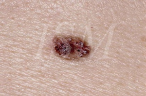
- Appearance evoking the classical basal cell carcinoma. This is the most common. Nevus then appears as a small smooth surfaced tumor translucent. Its coloration is variable, usually that of the normal skin, sometimes pale pink or gray. Consistency is much stronger than would be expected appearance but the base is not infiltrated. The size varies between 1 and 3 mm in diameter. If any of these lesions increased in size or starts to bleed or become covered with a crust, malignant transformation is suspected. Basal cell carcinomas appearing within the basal cell nevus syndrome can not be clearly distinguished from those appearing in an unaffected by this syndrome patient.The only characteristic element Gorlin syndrome is their occurrence in large numbers and at an early age and their location, which is not necessarily on sun-exposed areas. The clinical appearance of these tumors is highly variable ranging from flesh colored papules brown or ulcerative plaques. They can be mistaken for nevi or hemangiomas.Their size varies between 1 and 10 mm in diameter.
Histology:
The histological appearance of basal cell nevus is that of an intradermal proliferation of epithelial cells, forming well defined ranges, often with peripheral palisade sitting. The underlying stroma is rich in fibroblasts. The histological appearance of basal cell carcinoma patients Gorlin syndrome is not different from those found in non-syndromic patients. When nevi becomes basal cell carcinoma, one notices larger and clearer cells, increased number of mitoses, an inflammatory character of the infiltrate. It has a fibrous stroma. The lesion may become papullaire or stalked.Deep penetrations, ulcers or invasions can occur with lymphocytic infiltration. They can be pigmented in the mass or in the periphery.
Influence of the sun:
A study suggests that patients with basal cell nevus syndrome would present increased sensitivity to ultraviolet rays.This is supported by the fact that only 14% of patients with Gorlin syndrome in north-west England develop basal cell carcinomas, against 47% in Australia. It stresses the importance of prenatal diagnosis (when one parent is reached) or the earliest possible diagnosis (for cases appeared de novo) to limit UV exposure upon diagnosis positive.
Another study, carried reports that 88% of basal cell carcinomas in women and 86% among men in the general population occur on body areas exposed to the sun against 59% among women and 65% among male carriers of the basal cell nevus syndrome . He concluded that the anatomical distribution of lesions suggests that the sun is not an essential factor in the development of basal cell carcinomas.Their presence in greater numbers in exposed areas, however, suggests that the sun can exacerbate the development of basal cell carcinomas and must protect themselves. He did not report any significant correlation between the number of basal cell carcinomas and the total number of hours of exposure to sunlight.
Only about 40% of African Americans with Gorlin syndrome develop basal cell carcinomas against 90% of Caucasians. The study in 105 patients with Gorlin syndrome shows that only 38% of African Americans have one or more basal cell carcinomas against 97% of Caucasians. Moreover, Caucasians develop basal cell carcinomas more early. Indeed, approximately 50% of Caucasians develop their first carcinoma 21.5 years and 90% at 35 years against 20% and 40% at the same ages in African-Americans
The smallest frequency basal cell carcinomas in the group of African-Americans may be due to better protection against ultraviolet rays due to the pigmentation of their skin.
Influence of ionizing radiation:
Ionizing radiation has also been implicated in the development of these tumors. Indeed, patients undergoing radiotherapy treatments often have numerous basal cell carcinomas in the irradiated areas. They appear on average oscillating in a period between 6 months and 3 years after irradiation against an average of 21 years with a non patient with Gorlin syndrome.
It should be noted that one of the earliest signs of the disease is medulloblastoma. Now it is generally treated with radiotherapy. The risk of developing BCC in the irradiated area is strongly increased. In addition, these carcinomas are much more aggressive and therefore more difficult to treat. For these reasons, we must minimize the radiotherapy treatments and encourage surgical treatment.
Evolution:
Basal cell carcinomas appear as part Gorlin syndrome appear to be more aggressive, more difficult to treat. They can go up to cause the death of the patient by invasion (metastasis in the brain and lungs mainly) or resistance to treatment. Malignant transformation occurs mostly after puberty. The most aggressive tumors most often affect the eyelids and nose and cause tissue damage that could lead to a significant disfigurement of patients.
B / palmoplantar hyperkeratosis General:
They also met under the terms of "pits", holes or wells palmoplantar. 26 Frequency: Occurring at an early age, their presence may be an important aid to early diagnosis of basal cell nevus syndrome because they are present in approximately 65% to 80% of cases. They met with roughly the same frequency in all age groups. Palmar hyperkeratosis hands after immersion in water for a few minutes.
Clinical appearance:
They correspond to small holes that are punctiform defects in the stratum corneum, which is partially or totally absent. Rarely above 1 or 2 millimeters in diameter and 1 mm in depth, their background can be pink or, if dirt has accumulated on the inside, dark. These lesions are encountered only on the hands and feet and are asymptomatic.When the clinical diagnosis is difficult, the practitioner can immerse for a few minutes the hands and / or the patient's feet in warm water. The pits appear more clearly then.
Histology:
Histological examination of a palmar pits showed a small front proliferative basal cells. Although there is a cellular disorganization within this front, a crack between the front and the adjacent stroma was not observed. Mild vascular proliferation and fibrosis were observed in the adjacent dermis. istologique
In conclusion, palmoplantar pits are a characteristic manifestation Gorlin syndrome. They are an important aid to the diagnosis of this syndrome because of their high frequency of occurrence and early age of their occurrence. For these reasons, they are among the major diagnostic criteria for the disease, with basal cell carcinomas and odontogenic keratocysts.
The frequency of skin cysts in relation to the basal cell nevus syndrome is between 35 and 75% depending on the study. Epidermoid cysts usually occur on areas of the body pilleuses and rarely on non pilleuses areas such as the palms and soles of the feet. This typical location at the ends of the body would be a distinctive clinical manifestation of basal cell nevus syndrome.
This histological description is very similar to that of odontogenic keratocysts, a major diagnostic criteria of the basal cell nevus syndrome. These skin keratocyst histologically relatives of keratocysts of the jaws, may be a typical trademark of the basal cell nevus syndrome.
In conclusion, the discovery of skin cysts mainly located on the hands and feet, some have a strong resemblance histologically with odontogenic keratocysts, must be considered a "clinical pearls" that can lead us towards a diagnosis of Gorlin syndrome .
c / Other dermatologic manifestations
Other dermatologic manifestations are found cited in the literature, but due to their very low frequency of occurrence, are not clearly described. Patients may thus have lipomas, milia of grain, stains "latte" or Pendulums of molluscum. Lipoma located over the right scapula. Also note the presence of numerous cutaneous nevi. The mollusca Pendulums are fibrous tumors and flaccid skin. They may be planar and plated, the feel depressed, or, conversely, projections and even pedunculated. The milia is a rare disease of the skin characterized by the formation of small elevations bright translucent, the size of a pinhead produced by degeneration colloid (transformation of cells in a kind of jelly) of the upper layers the dermis.
2 / SKELETON EVENTS
A / brain calcifications envelopes
It is possible to meet in children physiological or pathological intracranial calcification. The former are very rare. The latter are more common and are generally described in association with tumors (such as medulloblastoma), neuro-cutaneous syndromes, infections and neurodegenerative disorders. In the general population, intracranial calcifications appear to average around 12 years, whereas in patients with Gorlin syndrome, they are visible from the first years of life. They can therefore be used as an early indicator for the diagnosis of basal cell nevus syndrome.They usually relate to false brain, which is part of the dura mater forming a central partition between two hemispheres. Their frequency in patients Gorlin syndrome proves quite important, since oscillates between 65 and 85% according to the authors. They represent the most frequent radiological sign. Calcifications of the tentorium are also encountered but much more rarely than on the wrong brain. The tent of the cerebellum is the part of the dura mater between the brain and the cerebellum. It is also possible to meet with agenesis of the corpus callosum but this anomaly remains rare.
B / Abnormal skull shape
Patients with basal cell nevus syndrome have abnormalities of the shape of the skull. These anomalies, although belonging to minor criteria of the disease, diagnosis of great interest since they are visible from birth. In some patients the skull is abnormally large prominence with frontal bossing and parietal-temporal. The brows are prominent. The hypertelorism (excessive spacing of the eyes) is common and prognathism is sometimes reported.The base of the nose has collapsed and expanded. A nasal septum deviation can also be observed A study of 105 patients with Gorlin syndrome, macrocephaly is observed with greater frequency (50% of cases) in these patients compared to the normal population. It is the same for hypertélorisme which is present in 42% of cases.
C / rib abnormalities
Although it belongs to minor criteria of the disease, costal abnormalities are present in approximately 42% of patients while their frequency in the population is very low. They are largely present and are mainly represented by: - bifidités - of agenesis - costal synostosis. The synostosis is a total weld of two adjacent bones. It is also possible to observe abnormal enlargement of the anterior or posterior end of the ribs. The third, fourth and fifth ribs are most commonly affected by these defects even if all the ribs can be achieved. Some authors like Kimonis decided to classify the costal anomalies among the major criteria of the disease for two reasons. The first is their high frequency (42%) in patients compared to the general population. The second is that these anomalies can be easily detected at birth and thus represent a major diagnostic element of the basal cell nevus syndrome in the pediatric population.Applying the criteria of Kimonis, an earlier diagnosis of the disease can be performed in a large number of patients.Indeed, other major criteria for the disease, such as odontogenic keratocysts or multiple basal cell carcinomas are not present at birth; They mostly appear during childhood or adolescence. Clinicians should be very attentive to minor clinical signs of the disease during childhood. The presence of Gorlin syndrome should be sought in children born with rib anomalies, cardiac fibroma, palate or cleft lip, polydactyly or macrocephaly.
D / vertebral anomalies
According to a study of 105 patients with Gorlin syndrome, cases of scoliosis were more frequently detected in how the group of people affected. However, this difference does not prove large enough to be meaningful. Sick individuals appear to have more severe scoliosis than the general population and most often located on sites of developmental abnormalities such as hemi-vertebrae fusions or vertebral bodies. These developmental anomalies are encountered in 31% of cases. Spina bifida cases have been reported but with a slightly different frequency than the general population. Spina bifida is a defect consisting in a crack of the solder posterior axis of the default spine ossification points on one or more vertebrae, through which are hernia in the form of a more or less large tumor, the meninges and sometimes the marrow with a variable amount of cerebrospinal fluid. Cervical and thoracic columns are most affected by this defect in patients Gorlin syndrome
E / Abnormal bone of the hand and foot
Polydactyly is not a very common deformity as it is encountered in only 3% of cases, however, it is easily detectable and present from birth. Its diagnosis is easy. Patients Gorlin syndrome may also have a shortening of the fourth and / or fifth metacarpal. These malformations are among the minor criteria of the disease because they are infrequent.
F / Other skeletal manifestations
Sprengel deformity: 11% of people with basal cell nevus syndrome have a congenital deformity Sprengel or elevation of the scapula. This anomaly involves moving in upward and inward of one or both scapula with deformation and sometimes attachment to the spine of the displaced bone. This increase is due to a failure of the normal descent position of the bone during fetal life.
3 / EYE EVENTS
The frequency of ocular involvement remains unclear. This is because they take up little space in the description Gorlin syndrome, as most published studies on this disease treat major criteria. Eye abnormalities that are not the subject of a mention, not a true description.
A / Congenital malformations of the globe
The main eye defects encountered are cataract, glaucoma and coloboma whose frequencies combined are estimated at less than 14% by Gorlin.
Cataract is an eye disorder leading to lens opacity or that of its capsule.
Glaucoma is an eye condition characterized by increased ocular pressure above 20 mm Hg. It is due to discomfort in the normal flow of aqueous humor.
Coloboma is a defect of the lens consisting of a peripheral notch, single or multiple.
The hypertelorism or excessive distance between the eyes, is also frequently met as it is part of the typical facial characteristics of basal cell nevus syndrome.
In addition to these anomalies, it is also possible to find cases of strabismus, congenital cataracts, microphthalmia or retinitis pigmentosa. Retinitis pigmentosa is a degenerative process of the retina, bilateral, family and hereditary. It appears in childhood and is characterized by a progressive loss of visual acuity and visual field constriction, leading eventually to blindness
B / Tumor globe process
The eyelids are frequently the seat of basal cell nevi at risk for malignant transformation. This transformation can be manifested by the establishment of a torpid ulceration (that is to say, which does not evolve and does not regress) and invasive or by infiltration in depth with lid mutilation. Basal cell carcinomas of the eyelids are generally more aggressive and harder to treat. Furthermore, their treatment can cause a significant facial mutilation for the patient.
Case chalazion have also been reported. Chalazion is a small eyelid tumor from chronic inflammation of a meibomian gland, sebaceous gland in the tarsal cartilage, constituting the framework of the eyelids.
4 / EVENTS NEUROLOGIC
A / Méduloblastomes Statistics:
In the general population, medulloblastomas are the most frequent brain tumors in childhood and affect about 20% of all such tumors. The average age of diagnosis is 6 years and 3 months and boys are affected to a greater extent than girls. Traditional treatment of these tumors consists of surgical excision combined with radiotherapy and chemotherapy. The survival rate at 5 years is between 60 and 80%. Approximately 5% of less than 5 years patients with medulloblastoma are achieved by Gorlin syndrome. This rate drops to 1 or 2% in the case of adults. The average age medulloblastoma diagnosis in patients is much earlier because it is 2.1 years.
For this reason, it is very important to think about the presence of medulloblastoma in the first years of life of children at risk of developing basal cell nevus syndrome.
Patients syndrome have a better survival rate than non-patients.
Treatment :
Surgical resection, supplemented by radiotherapy and / or chemotherapy treatment is generally proposed to eradicate medulloblastomas. In the case of Gorlin syndrome, the age of onset of these tumors is earlier; gold plus radiation therapy is practiced, the sooner the risk of long-term sequelae (such as growth disorders or endocrine dysfunction) is important. In addition, at the irradiated areas, the risk of developing BCC or meningiomas is greatly increased. These basal cell carcinomas prove to be much more aggressive and resistant to treatment.
For these reasons, treatment with radiation therapy in persons infected syndrome should be avoided whenever possible.
Medulloblastomas also meet the 42 chemotherapy treatments, this treatment modality should be favored, at least among the younger patients.
Some features Gorlin syndrome, present at an early age, such as mental retardation or calcifications of the falx, can be interpreted only as manifestations caused by the tumor. In addition, the most classic signs of the disease, such as skin lesions or maxillary keratocysts, appear only later.
For these reasons, the basal cell nevus syndrome can be difficult to identify at this age despite the presence of the tumor.
Some authors think that medulloblastoma should become one of the major criteria of the disease because it is part, with bone alterations, the earliest signs. Genetic investigations and rigorous clinical examinations must be offered, in search of a possible Gorlin syndrome, all patients with medulloblastoma before age 3 and their families.
B / Other neurological manifestations
Meningiomas:
Only 10 cases of basal naevomatoses associated meningiomas have been reported in the literature. The age of patients is between 18 and 64 years. There is no sexual predominance. These tumors appear to grow in radiation zones induced by the treatment of a previous medulloblastoma.
mental retardation :
Cases of mental retardation and learning difficulties have also been reported in approximately 5% of patients.
5 / OTHER GENERAL EVENTS
A / ovarian fibroids:
Girls and women affected by Gorlin syndrome can present ovarian fibroids. The fibroid is a benign tumors of the most common ovarian and represents approximately 20% of all ovarian tumors. In their initial report in 1964, Gorlin and Goltz have estimated that ovarian fibroids were present in 75% of women living with Gorlin syndrome.
A series of studies carried out recently showed a prevalence of between 14 and 24%. Age of reach average is 30.6 years, but may range from 16-45 years. Note however that some of these tumors were found in patients younger than 3.5 years. For this reason, patients syndrome must undergo their first ultrasound before the age of 13 years and have regular gynecological care. Ovarian fibroids grow slowly and an average size of 6 cm diameter even though some may go up to 30 cm in diameter.
It has been reported that ovarian fibroids encountered in Gorlin syndrome are more frequently bilateral and calcified than in the general population. In the majority of cases, these tumors are completely asymptomatic. They do not interfere with fertility and patients rarely undergo malignant transformation. The treatment of these tumors is surgical.
B / heart Fibroids:
Cases of cardiac fibroids have been reported in connection with the basal cell nevus syndrome but are rare. They are 3 to 5% of all patients with a cardiac fibroma. They represent only 5% of cardiac neoplasms. They appear in childhood and in 85% of cases in children under 10 years. Their favorite spot is the interventricular septum. These tumors are to be found quickly because they appear very early in life and can lead up to a patient's early death.
C / hypogonadism in men:
Men with by Gorlin syndrome can present very rarely hypogonadism. Hypogonadism is the condition of a subject whose gonads have insufficient internal secretion.
6 / EVENTS ODONTOSTOMATOLOGIQUES
A. odontogenic keratocysts
1. General
Described for the first time by Philipsen in 1956, is a odontogenic keratocyst, according to the World Health Organization or WHO, "a benign uni- or multi-celled, intraosseous odontogenic origin, with a characteristic layer stratified squamous epithelium parakératinisé and potential for aggressive behavior and infiltrating. " WHO also recommends the term "tumor" as this better reflects the nature of neoplastic (aggressive behavior, major expansion, tendency to relapse ...). The term primordial cyst has long been used as a synonym of the term keratocyst, maintaining confusion. Indeed, all the primordial cysts are not keratocysts since they do not all keratinization.Conversely, all keratocysts are not essential cysts in so far as they do not always develop in place of a tooth.
The term epidermoid cyst is, meanwhile, a synonym. They represent about 11% of all cysts of the jaws. They rank third, after the radicular and follicular cysts. They appear three times more frequently in the mandible than in the maxilla.
The odontogenic keratocysts occur in all age groups, although a cumulative frequency is observed between ten and forty years in the normal population. By cons, in patients Gorlin syndrome, the peak frequency is earlier and is therefore in the first decade of life. These cysts thus constitute an early symptom of the disease. This confirms the place of choice of the dentist in the early detection of basal cell nevus syndrome. They are most of the time discovered by chance during a standard radiography. The percentage of asymptomatic patients varies depending on the study from 34 to 50%. They also may be revealed by the appearance of symptoms such as swelling, pain most often; but trismus, cellulitis, a delayed eruption or dental malposition among others ...
Note that all these symptoms are not specific of odontogenic keratocysts. The presence of keratocyst odontogenic in a young patient or the appearance of recurrent or multiple keratocysts should alert the practitioner and towards a possible diagnosis of Gorlin syndrome. Approximately 5% of keratocysts are associated with this syndrome. They are found in 80% of patients with Gorlin syndrome when they are present in only 5-7% of the general population.There are several theories to explain the etiology of odontogenic keratocysts.
Some odontogenic keratocysts would develop from dental plates.
Indeed, following the formation of dental germs, dental strips dissolve in both jaws. During this phenomenon, islets and epithelial cell clusters can persist in the connective tissue. It would epithelial these residues, which upon activation by yet unknown factors are the cause of the formation of keratocysts. According to other, they emanate from the stellate reticulum of the enamel organ or the basal layer of the epithelium of the oral mucosa. Indeed, this other theory assumes that the ramifications of the basal layer are the cause of the formation of keratocysts. One argument supporting this theory is that observed relapses of these cysts even when surgical resection of the affected bone segments. This assumes that the origin of the recurrence is outside of this bone and probably in adjacent soft tissue. It should be noted that these two theories are not incompatible since both the dental lamina as oral mucosa are responsible for ectodermal tissues.
2. Features
The histological diagnosis of odontogenic keratocyst can be done by the histolopathologique analysis of it.Radiological examination can certainly guide the practitioner but in no case make a definitive diagnosis. This examination consists of the content analysis and cystic walls is obtained after complete resection of the cyst or after taking a fragment of it during his or her marsupialisation drainage. The main histological characteristics listed by most writers are those of Pinborg and Hansen in 1966:
- Squamous epithelium lining usually very fine and uniform in thickness, with little or no taste buds and with a strong mitotic activity;
- A well defined basal cell layer. The cells that compose it are cubic or cylindrical shapes and arranged most often in a palisade arrangement;
- A thin layer, from four to eight cell layers of spiny cells frequently presenting an intracellular edema;
- Keratinization is, usually, parakeratotic (presence of nuclei). It occurs in 83-97% of keratocysts. Sometimes it is possible to meet a keratinization orthokeratosic kind (no nuclei) or both para- and orthokeratosic. For some authors like Martin, the orthokeratosic kind should be individualized because it would be less aggressive and less recurrent as parakeratotic kind;
- Keratin layer is often wavy. The only keratinization is not a specific criterion keratocyst because other odontogenic cysts can produce keratin.
- The fibrous cyst wall is usually thin and does not usually inflammation;
- A thin shell conjunctiva often containing epithelial islands or cysts "girls" isolated periphery. The luminal contents of the cyst has a liquid aspect, pale or creamy yellow, whiter, thicker. There is a presence of keratin, but in varying amounts. In case of inflammatory reaction is found in this content cholesterol crystals (being in the form of shiny flakes), hyaline body, polynuclear and bacterial clumps. In addition, when the cyst wall is inflamed, the epithelium is thickened and gradually turns into a non-keratinized epithelium quite similar to that odontogenic inflammatory commonplace cysts. FNA is also an important test for the diagnosis of odontogenic keratocyst.
, In fact, the presence of this keratin in FNA is a diagnostic aid element (but that alone is not enough to make a definitive diagnosis because other cysts such as dentigerous cysts, primary root or have an epithelium surface which can be keratinized).
However, as described above, the presence of inflammatory reaction induced progressive loss of parakératinisation of keratocysts and thus can sometimes prevent correct cytological diagnosis. One of the other elements of FNAB for referral to a diagnostic keratocyst is the estimation of the protein content of the cyst.
When this content is measured at least 4 grams of protein per 100 ml of liquid, the diagnostic keratocyst is then relatively safe. For this measurement, the presence of inflammation also has a bad influence.
Indeed, it tends to increase the protein content and therefore participated in the creation of false negatives. There are also methods to realize the diagnosis before turning to surgery. These techniques are not yet common practice.The first is the search for KAC antigen (keratocyst antigen) in the cyst fluid. This presence confirms the diagnosis.The second is the determination of the immunohistochemical expression of cytokeratin 10.
Indeed, it is a marker of the keratinized epithelia. It is found in about 50% of odontogenic keratocysts, but also in 10% of radicular cysts and 3% of dentigerous cysts. However the study of the expression of cytokeratin this would not be in the keratocysts insightful as these lesions seem to lose the expression after decompression.
3. Clinical and radiological features
Location:
The keratocyst odontogenic seat more often in the mandible in the maxilla. Indeed, it occurs three times more often in the mandible, especially at angle level and the ramus, followed by locations in the area first and second molars and more anterior locations.
At the headquarters of the maxillary keratocyst is a little earlier in the prémolomolaire region and the canine and exceptionally the anterior region.
Age of onset: The odontogenic keratocysts associated with Gorlin syndrome develops, in general, during the first decade of life and reach their peak incidence between 20 and 30 years, unlike not associated with this syndrome and keratocysts are discovered later, during the fourth decade (average age 37 years).
Because of this early development, they constitute one of the first noticeable symptoms of the disease and confirm the place of choice for dentistry in the diagnosis of this syndrome.
Size:
Their volume is more variable, ranging from a few millimeters to a flooding of a hemi-mandible (18). Because they are often discovered later, during a routine X-ray examination, it is not uncommon that they have already reached a considerable volume. Their average length of 5 centimeters. (34)
Sex ratio:
In the normal population men are affected more often than women with a sex ratio of 2/1. By cons in patients with basal cell nevus syndrome, the difference between the sexes does not exist and it even seems that women are affected slightly more often than men. (5) Symptoms: The majority of odontogenic keratocysts is free of any symptoms. Indeed, 34 to 50% are asymptomatic and thus discovered only during a dental or radiological examination routine. When symptoms exist, they occur most often by the appearance of an arch or progressive bone swelling can be painful.
The inflammatory phenomena are then not uncommon and are characterized by the occurrence of abscesses, cellulitis or even sometimes accompanied fistulisations a lockjaw. Outside of these phenomena, the keratocyst can be discovered because of misalignments, dental malocclusions or delays it may cause rash and which will challenge the practitioner.
It should be noted that all these manifestations are not at all specific to odontogenic keratocysts and will require a radiological examination and especially histopathological order to confirm the diagnosis.
Growth:
One of the main clinical characteristics of odontogenic keratocysts is their strong growth potential. This is even one of the criteria that led WHO to consider them as benign neoplasms. Various theories have been put forward to explain this characteristic:
- A first explanation would be the existence of a collagenase enzyme inside the keratocyst. She would be responsible for a collagenolytic activity which explains this strong growth potential;
- Another theory is the existence of bone resorption induced by prostaglandin E and F secreted by the cyst;
- Finally, greater osmolarity of cystic fluid and an increased mitotic power at the cyst wall could also be responsible for this strong growth. Extending these cysts is within the bone substituting cancellous bone rather than extending laterally as other cysts towards the cortical and periosteal regions.
When keratocysts are bulky, they are molded on the edges of the bones and teeth, cortical 53 (usually external) is then thinned, sometimes perforated. Cases of mandibular and / or major facial deformities fractures have been reported but are however very rare. The noble elements such as the neurovascular bundle of the inferior alveolar canal are, in turn, always respected, but can be turned back (and very rarely cause paraesthesia).
Recurrence, recurrence mechanism :
The odontogenic keratocysts also have a very high recurrence rate. This is one of their main clinical feature. This rate is even higher in patients with basal cell nevus syndrome and is around 82% against 25-60% in healthy patients (33). There are three mechanisms explaining the reasons for this high rate of recidivism:
- Incomplete excision of the peripheries of the cyst or because of a very thin and friable cyst wall, either due to a perforation of the cortical bone with the cyst adhesion to adjacent soft tissue;
- The occurrence of odontogenic keratocyst from a satellite cyst, a microcyst odontogenic remnants forgotten or after surgery;
- The development of a new keratocyst odontogenic in an adjacent area and which is interpreted as a recurrence while it is a de novo formation having no connection with the original lesion. According to other authors, the recurrence rate is also influenced by:
- The size, location (Ramus), the morphology of the lesion (especially multiloculaire) that make treatment more difficult and surgery
- The choice of surgical technique (conservative or aggressive) Most recurrences are found in the 5-7 years of the first support.
However, cases have been reported more than 10 years after treatment. 25% of recurrent cases are located from 9 years and older. 54 An essential aspect of odontogenic keratocysts treatment is monitored in the long term (ten years seems ideal to be able to diagnose almost all recurrences at an early stage). Some features of odontogenic keratocysts have brought WHO to reclassify to as tumors or benign neoplasms rather than cysts: - first behavior: these cysts are locally very destructive, invasive and have a high recurrence rate; - Then their histopathology: studies have shown that the basal layer buds in the connective tissue. WHO further noted that mitoses figures are frequently found in the supra-basal layer; epithelium keratocysts a mitotic index increased relative to the other jaw cysts (but similar to ameloblastoma); - To finish the genetics: the PTCH gene is a tumor suppressor gene located on chromosome 9q22.3-q31. This gene is responsible for both the basal cell nevus syndrome but also responsible for the formation of isolated odontogenic keratocysts. Normally PTCH codes for a transmembrane protein that forms a signal receiver with the oncogene SMO (smoothened) for ligand SHH (sonic hedgehog). SHH receptor activates this when there is an inhibition of the growth signal of different tissues and proliferation. It is also involved in the regulation of lipid metabolism. By cons if one loses the operating PTCH, we will have a cell proliferation effect (and therefore development of cysts and tumors).
Standard radiological aspect:
Very often asymptomatic, most keratocysts are discovered during a standard radiological examination. This examination in no case make a definitive diagnosis. It allows an orientation of the diagnosis can only be confirmed by histopathological analysis. The most common X-ray appearance of odontogenic keratocyst is the mono (usually) or polygéodes radiolucent, round or oval and with thin internal partitions.
In the maxilla, overlays with sinus or nasal cavities can make the identification of these features much more difficult.When keratocyst is very extensive, thinning and cortical bone deformation is possible. It can develop on any hemi-maxilla; the condyles are always respected.
The latter can also wrap an unevolved adjacent tooth (included), a supernumerary tooth or a odontome and cause the displacement or, very rarely, the root resorption of other teeth located on contact. Peripheral noble elements (nerves ...) are not destroyed but sent back by the cyst. The main differential radiological diagnosis arises with ameloblastoma, dentigerous cysts then some.
4. Evolution
The cases of malignant transformation of odontogenic keratocysts in squamous cell carcinomas are very rare. Only 12 cases have been reported in the literature to date. This possibility should be considered when there are multiple recurrences, a very aggressive behavior of the lesion as well as a failure of both the conservative treatments and aggressive treatments. It is often observed in elderly patients. Case ameloblastic transformations are reported but also very rare
. 5. Differential Diagnosis
The differential diagnosis of odontogenic keratocyst must be done with three other cysts mainly:
- with ameloblastoma:
This cyst is most frequent in males and has its peak incidence in the fourth and fifth decades. 80% of ameloblastomas are located in the mandible and especially at angle level and the ramus. Maxillary localizations are rare.
From a clinical standpoint, ameloblastoma is characterized by unlimited growth and a high recurrence rate. These lesions often have a large enough volume. Generally asymptomatic, it will be discovered only by chance of a radiographic examination. Otherwise, it can be revealed by an infectious episode or by movements and dental anomalies evolution.
From a radiological point of view, ameloblastoma has mostly a polygéodique aspect "soap bubble", regular borders and with internal partitions. Geodes are well demarcated, round or oval, of varying sizes. The noble elements are repressed, not destroyed. There are so many similarities with the keratocyst, making the differential diagnosis impossible with the only standard clinical and radiographic findings. The differential diagnosis will therefore mainly by histological examination but also by the CT scan.
- with a dentigerous cyst:
This cyst met more frequently in men has its peak incidence in the second and third decades. It essentially concerned the third lower molar and the upper canine and the lower second premolar.
Mostly free of symptoms, the dentigerous cyst adheres to the neck of an impacted tooth and surrounds the crown.His discovery is most often during a routine X-ray examination or may be motivated by unexplained tooth movement.
The radiographic appearance is that of a well-demarcated osteolysis, monogéodique and developed around the wreath or crown of impacted tooth and totally built. The lesion can become quite large and invade the ascending branch of the mandible. It is more difficult to perceive radiologically inserting it in the neck of the impacted tooth and therefore the differentiating a keratocyst.
In addition, it is possible to observe a root resorption of teeth adjacent to the lesion; which is much rarer for odontogenic keratocysts and is the main distinctive radiological sign of these two entities.
- With a follicular cyst:
This cyst is a variety of cyst Dentigerous but which develops in contact with dental follicles; that is to say, teeth incompletely formed. Its discovery can be done either by means of a radiograph motivated by a delayed eruption of a permanent tooth; either during an infectious episode (usually in the premolar region). Other features are identical to those of Dentigerous cyst.
- With a banal inflammatory cyst or dental radiculo:
These lesions are most frequent maxillary. They preferentially occur in males in the anterior maxillary or the horizontal branch of the mandible. Their peak incidence is between the third and fourth decades. Similarly lesions seen previously, the radicular cyst is usually asymptomatic and radiological routine discovery. It can however be revealed in infectious episodes characterized by bulging, swelling, pus beside a decayed tooth, necrotic or incompletely processed. These cysts are slow growing and may, in the absence of clinical signs, expand considerably.
Their radiographic appearance is that of a monogéode, rounded or oval, in connection with the apex of a tooth mortified and surrounded by a border bone condensation (which may disappear during inflammatory attacks). They may, in case of significant size, cause root resorption of the adjacent roots. 59
The differential diagnosis may also land with other cysts such as the odontogenic fibroma, the odontogenic myxoma, the solitary cyst, bone angioma or the aneurysmal cyst. 60 B.
B / Other events odontostomatologiques
Other Dental odonto- events are also encountered in the basal cell nevus syndrome but with a greater disparity.Indeed, they are not always present, and when they are, it is with a frequency much lower than the odontogenic keratocysts which are, themselves, one of the major criteria of the disease. They are often directly related to the development of keratocysts and may represent one of the first call signs to highlight them (and therefore lead to the diagnosis of basal cell nevus syndrome)
a / Inclusions or dental malposition
They mainly concern the permanent dentition. It is possible to observe a persistence of primary teeth when the final tooth is retained by the cyst, which prevents its eruption. Studies have demonstrated that, depending on the time development of the cyst, the consequences dental level are different. Indeed, when the keratocyst develops when root construction, deformation was observed in the roots such as curvatures or bends bayonet.
If it develops early in juxtaposition to a dental element formation, may cause the tearing of the roots. By cons, if the cyst is formed after tooth eruption, he may be responsible for travel and dental malposition but in any case it can not be responsible for root resorption or pulp mortification.
b / mandibular prognathism
An estimated 25% of cases. It may be either a true prognathism or a pseudoprognathisme linked to a brachymaxillie, or a combination of both. 61 Their support will be through orthognathic surgery and orthodontics.Mandibular prognathism in a young patient with Gorlin's syndrome (15)
c / lip and palate clefts
The cleft lip and / or cleft palate are found in about 5-7% of cases of basal cell nevus syndrome. A literature review conducted by Lambrecht shows that of 719 people affected by Gorlin syndrome, orofacial have 61 slot, 8.5% of patients. These people can be divided into two groups depending on whether the slot is labial, alveolar and palate (in 49 cases) or only cleft (for 13 cases)
The frequency of these malformations is not the same between the general population and patients with basal cell nevus syndrome
.Indeed, in the general population, the incidence of labial slits, alveolar and palate is that of only 1/800 and 1/2500 against cleft palate respectively 1 / 14.7 and 1 / 55.3 of patients Gorlin syndrome. This abnormality may be one of the first indications of the presence of a Gorlin syndrome in a newborn.
d ./ paresthesia
They are also linked to the development of keratocysts and occur when the cyst reaches a significant size if it compresses or represses a nervous element and causes paresthesia. It can also be a postoperative complication of removal of a large cyst with boundaries near the nerve.
e / The améloblastome
The ameloblastomas are rarely encountered in patients Gorlin syndrome since few cases have been reported in the literature. Eslami shows the case of a 68 year old patient achieved by Gorlin syndrome and having a ameloblastoma.
While for almost all patients affected by this disease, the first symptoms appear at an early age, usually before age 35, the first clinical signs of the patient began to appear much later.
Indeed, it presented its first basal cell carcinoma at the age of 50 years and multiple keratocysts from 68 years only.Element interesting, we Eslami noted that 3 of the 4 cases of basal cell nevus syndrome associated with ameloblastoma recorded in English literature, also presented their first symptoms at an older age than the majority of cases of basal cell nevus syndrome.
The average age for these cases is 47.6 years. This discovery indicates that patients with basal cell nevus syndrome at a later age are more at risk of developing ameloblastomas.
As seen above, odontogenic keratocysts and ameloblastomas are, both, benign neoplasms with a high recurrence rate and great potential of bone destruction.
However, ameloblastoma is more persistent and more infiltrating, requiring an even more radical surgical resection.
